Sub-Total: $0.00 USD
Have questions about this assay kit?
Human G-CSFR Reporter Assay Kit
Product Description and Product Data
This is an all-inclusive cell-based luciferase reporter assay kit targeting the the Human Granulocyte Colony-Stimulating Factor Receptor (G-CSFR). INDIGO’s G-CSFR reporter assay utilizes proprietary mammalian cells that have been engineered to provide constitutive expression of the G-CSFR. In addition to G-CSFR Reporter Cells, this kit provides two optimized media for use during cell culture and in diluting the user’s test samples, a reference agonist, Luciferase Detection Reagent, and a cell culture-ready assay plate. The principal application of this assay is in the screening of test samples to quantify any functional activity, either agonist or antagonist, that they may exert against G-CSFR. This kit provides researchers with clear, reproducible results, exceptional cell viability post-thaw, and consistent results lot to lot. Kits must be stored at -80C. Do not store in liquid nitrogen. Note: reporter cells cannot be refrozen or maintained in extended culture.
Features
Clear, Reproducible Results
- All-Inclusive Assay Systems
- Exceptional Cell Viability Post-Thaw
- Consistent Results Lot to Lot
Product Specifications
| Target Type | Growth Factor Receptor | ||
| Species | Human | ||
| Receptor Form | Hybrid | ||
| Assay Mode | Agonist, Antagonist | ||
| Kit Components |
| ||
| Shelf Life | 6 months | ||
| Shipping Requirements | Dry Ice | ||
| Storage temperature | -80C |
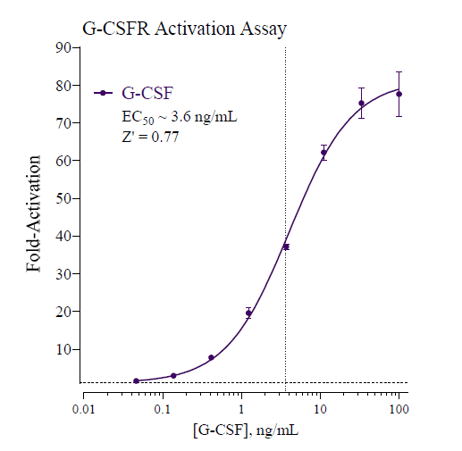
Data
Target Background
Granulocyte colony-stimulating factor (G-CSF) is a hematopoietic cytokine that regulates the viability, proliferation, and differentiation of granulocyte precursors and the function of neutrophils by signaling through the homo-dimeric granulocyte stimulating factor receptor (G-CSFR; also known as CSF3R). G-CSFR is a single transmembrane protein that has no intrinsic tyrosine kinase activity; however, upon ligand binding the receptor undergoes a conformational change leading to the activation of several downstream pathways. G-CSFR is predominantly expressed on neutrophils throughout all stages of maturation but is also present on myeloid progenitors and endothelial cells.
In chemotherapy-induced neutropenia, the bone marrow reserve of granulocytes is decreased. Exogenous G-CSF treatment can accelerate proliferation and differentiation of progenitor cells, aiding neutrophil replenishment.
G-CSF is known to play an important role in cancer development and progression. Acute myeloid leukemia (AML) and atypical chronic myelogenous leukemia (aCML) have been disorders directly related to G-CSFR mutations. In addition, G-CSF and G-CSFR are highly expressed in 90% of human gastric and colon tumors.
G-CSF is also known to contribute to chronic inflammatory diseases by stimulating the activation and migration of myeloid cells to inflammation sites. G-CSF and G-CSFR deficient mice are profoundly protected in several models of rheumatoid arthritis (RA), and antibody blockade of G-CSF has been shown to protect against disease3. Consequently, GCSF and its specific receptor, G-CSFR, command considerable interest in therapeutics development and drug safety screening.
Citations
Also available as a service