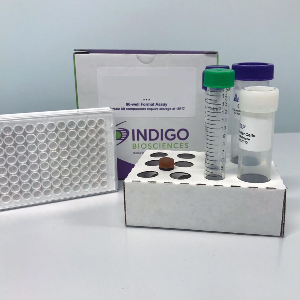
Product Description and Product Data
This is an all-inclusive cell-based luciferase reporter assay kit targeting the the Human Granulocyte-Macrophage Colony-Stimulating Factor Receptor (GM-CSFR). INDIGO’s GM-CSFR reporter assay utilizes proprietary mammalian cells that have been engineered to provide constitutive expression of the GM-CSFR. In addition to GM-CSFR Reporter Cells, this kit provides two optimized media for use during cell culture and in diluting the user’s test samples, a reference agonist, Luciferase Detection Reagent, and a cell culture-ready assay plate. The principal application of this assay is in the screening of test samples to quantify any functional activity, either agonist or antagonist, that they may exert against GM-CSFR. This kit provides researchers with clear, reproducible results, exceptional cell viability post-thaw, and consistent results lot to lot. Kits must be stored at -80C. Do not store in liquid nitrogen. Note: reporter cells cannot be refrozen or maintained in extended culture.
Features
Clear, Reproducible Results
- All-Inclusive Assay Systems
- Exceptional Cell Viability Post-Thaw
- Consistent Results Lot to Lot
Product Specifications
| Target Type | Growth Factor Receptor | ||
| Species | Human | ||
| Receptor Form | Hybrid | ||
| Assay Mode | Agonist, Antagonist | ||
| Kit Components |
| ||
| Shelf Life | 6 months | ||
| Shipping Requirements | Dry Ice | ||
| Storage temperature | -80C |
Data
Target Background
The glycoprotein, granulocyte-macrophage colony-stimulating factor (GM-CSF) was originally defined as a growth factor that induces the differentiation and proliferation of myeloid progenitors in response to stress, infections, and cancers.
The activity of GM-CSF is mediated through binding interactions with the GM-CSF Receptor (GM-CSFR; also known as CSF2R). GM-CSFR is expressed in myeloid cells and some non-hematopoietic cells as a heterodimer comprising a ligand-specific alpha subunit and a beta subunit. The beta subunit is also shared with the heteromeric IL-3 and IL-5 receptors.
GM-CSF is known to play an important role in cancer development and progression. GM-CSF stimulates the production and maturation of neutrophils, macrophages and dendritic cells that mediate the initial host responses to cancers. However, a fine balance exists. Too little GM-CSF prevents the appropriate production of innate immune cells and subsequent activation of adaptive anti-cancer immune responses. Conversely, too much GM-CSF production can exhaust immune cells and promote cancer growth.
GM-CSF is also commonly described as a cytokine, and is known to contribute to chronic inflammatory diseases by stimulating the activation and migration of myeloid cells to inflammation sites. An imbalance in GM-CSF production and signaling has been associated with autoimmune diseases like multiple sclerosis (MS), rheumatoid arthritis (RA), myasthenia gravis (MG), inflammatory bowel disease (IBD) and systemic lupus erythematosus (SLE). Consequently, GM-CSF and its specific receptor, GM-CSFR, command considerable interest in therapeutics development and drug safety screening.
Product Documentation
Also available as a service
