Sub-Total: $0.00 USD
Have questions about this assay kit?
Human M-CSFR Reporter Assay Kit
Product Description and Product Data
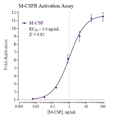
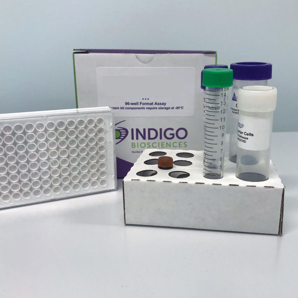
This is an all-inclusive cell-based luciferase reporter assay kit targeting the the Human Macrophage Colony-Stimulating Factor Receptor (M-CSFR). INDIGO’s M-CSFR reporter assay utilizes proprietary mammalian cells that have been engineered to provide constitutive expression of the M-CSFR. In addition to M-CSFR Reporter Cells, this kit provides two optimized media for use during cell culture and in diluting the user’s test samples, a reference agonist, Luciferase Detection Reagent, and a cell culture-ready assay plate. The principal application of this assay is in the screening of test samples to quantify any functional activity, either agonist or antagonist, that they may exert against M-CSFR. This kit provides researchers with clear, reproducible results, exceptional cell viability post-thaw, and consistent results lot to lot. Kits must be stored at -80C. Do not store in liquid nitrogen. Note: reporter cells cannot be refrozen or maintained in extended culture.
Features
Clear, Reproducible Results
- All-Inclusive Assay Systems
- Exceptional Cell Viability Post-Thaw
- Consistent Results Lot to Lot
Product Specifications
| Target Type | Cytokine Receptor, Growth Factor Receptor | ||
| Species | Human | ||
| Receptor Form | Hybrid | ||
| Assay Mode | Agonist, Antagonist | ||
| Kit Components |
| ||
| Shelf Life | 6 months | ||
| Shipping Requirements | Dry Ice | ||
| Storage temperature | -80C |
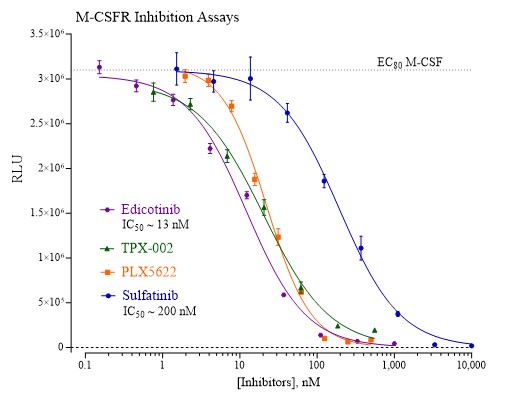
Data
Target Background
Macrophage colony-stimulating factor (M-CSF) is a cytokine expressed in a wide range of cells and tissues. It stimulates progenitor cells from bone marrow and plays a role in the development, proliferation, and maintenance of phagocytes like monocytes, dendritic cells and microglia.
The activity of M-CSF is mediated through binding interactions with the M-CSF Receptor (M-CSFR; also known as CSF1R). M-CSF/M-CSFR activity is involved in various pathologies such as ovarian cancer, breast cancer, rheumatoid arthritis, and cutaneous lupus. Recent phase II clinical trials have indicated that monoclonal antibody or small molecule antagonists targeting M-CSFR have reduced the inflammatory response often associated with rheumatoid arthritis.
In the brain, M-CSF is secreted by neurons, astrocytes and microglia and is involved in brain development. M-CSF-deficient animals have severe brain deficits with abnormalities associated with the cerebral cortex. Other phenotypic traits associated with M-CSF deficiency include reduced body weight and skeletal defects. Additionally, in many neurodegenerative diseases, microglia are becoming a key target for therapeutic purposes as they eliminate toxic elements from the brain and set the conditions for repair and remyelination. Low level M-CSF/M-CSFR expression or receptor inhibition in microglia has been associated with pre-symptomatic Alzheimer’s disease (AD), Parkinson’s disease (PD), multiple sclerosis (MS) and frontotemporal dementia. Consequently, M-CSF and its specific receptor, M-CSFR, command considerable interest in therapeutics development and drug safety screening.
Product Documentation
Citations
Also available as a service