Sub-Total: $0.00 USD

Have questions about this assay kit?
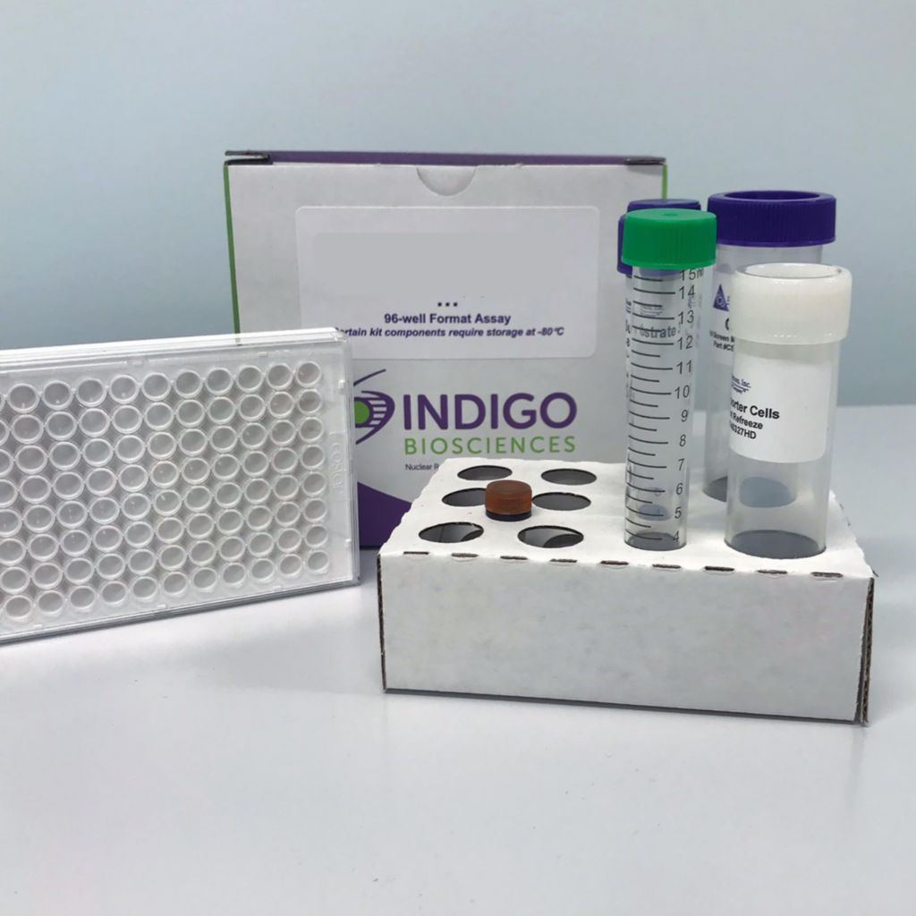
Human EGFR1 Reporter Assay Kit
Product Description and Product Data
This is an all-inclusive cell-based luciferase reporter assay kit targeting the Human Epidermal Growth Factor Receptor 1 (EGFR1). INDIGO’s EGFR1 Reporter Cells include the luciferase reporter gene functionally linked to a EGFR1 -responsive promoter. In addition to EGFR1 Reporter Cells, this kit provides two optimized media for use during cell culture and in diluting the user’s test samples, a reference agonist, Luciferase Detection Reagent, and a cell culture-ready assay plate. The principal application of this assay is in the screening of test samples to quantify any functional activity, either agonist or antagonist, that they may exert against human EGFR1. This kit provides researchers with clear, reproducible results, exceptional cell viability post-thaw, and consistent results lot to lot. Kits must be stored at -80C. Do not store in liquid nitrogen. Note: reporter cells cannot be refrozen or maintained in extended culture.
Features
Ready to Use Upon Receipt
- Includes All Needed Components
- Contains Transfected Reporter Cells
- Eliminates Cell Licensing Fees
- Clear, Reproducible Results
- Consistent Results Lot to Lot
Product Specifications
| Target Type | Growth Factor Receptor | ||
| Species | Human | ||
| Receptor Form | Native | ||
| Assay Mode | Antagonist | ||
| Kit Components |
| ||
| Shelf Life | 6 months | ||
| Orthologs Available | No | ||
| Shipping Requirements | Dry Ice | ||
| Storage temperature | -80C |
Data

Target Background
EGFR1 is a single-pass transmembrane receptor, one of four members of the receptor tyrosine kinase (RTK) family. Binding interactions with extra-cellular signaling peptides such a epidermal growth factor (EGF), transforming growth factor alpha (TGFα), or amphiregulan lead to receptor dimerization and auto-phosphorylation and activation of associated intracellular signaling proteins. Interestingly, EGF Receptors demonstrate two alternative signal processing modes: one via the membrane-bound receptor and signaling at cell surface; and the other signaling through internalized receptors, nuclear translocation, and co-association with activated transcription factors to interact directly with target gene promoter sequences.
Activated EGF Receptors are known to signal through several different pathways, including those mediated by Ras, PI3K, PLC-γ, and JAK, culminating in the activation of specific transcription factors and the induction of respective target genes. Phosphorylation and activation of the transcription factor STAT3 is one prominent pathway utilized by EGFR1, and it is the signaling mechanism exploited by the reporter cells in the INDIGO kit.
INDIGO’s reporter cells contain the luciferase gene functionally linked to an upstream minimal promoter and tandem STAT3 genetic response element (GRE) sequences. Activated, dimeric STAT3P (or STAT3P associated with nuclear EGFR1P) bind the STAT3 GREs to initiate the formation of a complete transcription complex that drives Luc expression. Quantifying relative changes in luciferase activity in the treated reporter cells relative to the untreated cells provides a sensitive, dose-dependent surrogate measure of drug- or antibody-induced changes in EGFR1 activity.
Considering their significant role in rapid cell proliferation, and their involvement in the progression of many types of cancers, EGFR continues to command much interest as a target for the development of novel, specific, and predominantly inhibitory drugs and antibodies. Accordingly, the primary application of this EGFR1 Reporter Assay is to screen test materials for any functional activity, either agonist or inhibitory, that they may exert against the EGFR1.
Citations
Also available as a service