View the slide viewer

Have questions about this assay kit?

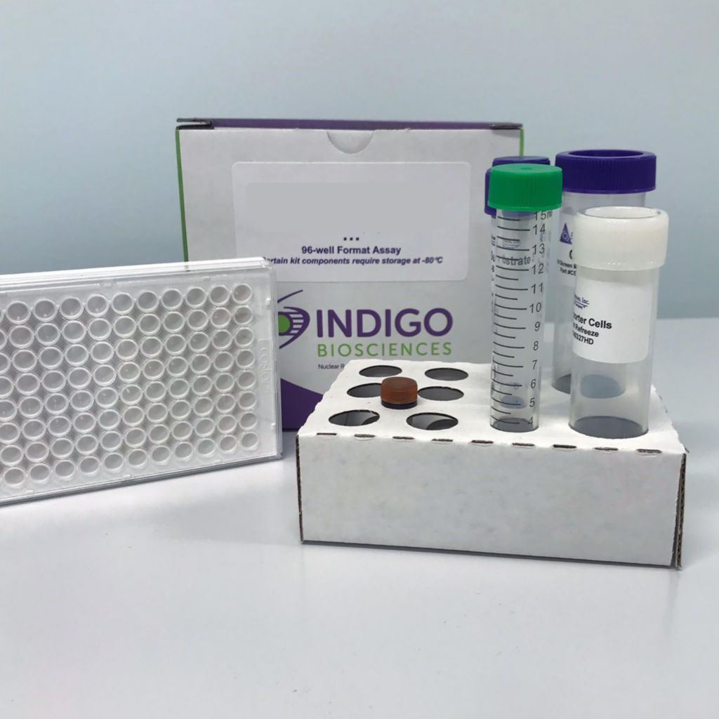
Human RARα Reporter Assay Kit

SIZE SKU PRICE
1 x-96 well format assays
3 x-32 assays in-96 well format
1 x-384 well format assays
SIZE SKU
1 x-96 well format assays
3 x-32 assays in-96 well format
1 x-384 well format assays

Product Description and Product Data

This is an all-inclusive cell-based luciferase reporter assay kit targeting the Human Retinoic Acid Receptor alpha (RARa). INDIGO’s RAR Alpha reporter assay utilizes proprietary mammalian cells that have been engineered to provide constitutive expression of the RAR Alpha. In addition to RAR Alpha Reporter Cells, this kit provides two optimized media for use during cell culture and in diluting the user’s test samples, a reference agonist, Luciferase Detection Reagent, and a cell culture-ready assay plate. The principal application of this assay is in the screening of test samples to quantify any functional activity, either agonist or antagonist, that they may exert against human RAR Alpha. This kit provides researchers with clear, reproducible results, exceptional cell viability post-thaw, and consistent results lot to lot. Kits must be stored at -80C. Do not store in liquid nitrogen. Note: reporter cells cannot be refrozen or maintained in extended culture.

Features

  • Ready to Use Upon Receipt

  • Includes All Needed Components
  • Contains Transfected Reporter Cells
  • Eliminates Cell Licensing Fees
  • Clear, Reproducible Results
  • Consistent Results Lot to Lot

Product Specifications

Target TypeNuclear Hormone Receptor
SpeciesHuman
Receptor FormHybrid
Assay ModeAgonist, Antagonist
Kit Components
  • RARa Reporter Cells
  • Cell Recovery Medium (CRM)
  • Compound Screening Medium (CSM)
  • 9-cis Retinoic Acid, (ref. agonist; in DMSO)
  • Detection Substrate
  • Detection Buffer
  • White, sterile, cell-culture ready assay plate
Shelf Life6 months
Orthologs AvailableYes
Shipping RequirementsDry Ice
Storage temperature-80C

Data

Agonist dose-response analyses of the RARα Assay. Validation of the RARα Assay was performed using manual dispensing and following the protocol described in the assayTechnical Manual, using reference agonists 9-cis-Retinoic Acid (9-cis-RA; provided), AM80 (Tocris), AM580 (Tocris) and BMS753 (Tocris). In addition, to assess the level of background signal contributed by non-specific factors that may cause activation of the luciferase reporter gene, “Mock” reporter cells were specially prepared to contain only the luciferase reporter vector (mock reporter cells are not provided with assay kits). Final assay concentrations of agonist treatment media ranged between 2.5 µM and 2.5 pM, and included a 'no-treatment' control (n ≥ 6 / treatment; highest [DMSO] ≤ 0.025% f.c.. APPENDIX 1 in the tech manual describes an abbreviated 8-point dilution scheme.) Mock Reporter Cells were identically treated with 9-cis-RA. Luminescence was quantified using a GloMax-Multi+ plate-reading luminometer (Promega Corp.). Average Relative Light Units (RLU) and their respective values of Standard Deviation (SD), Coefficient of Variation (CV), and Signal-to-Background (S/B) were determined for each treatment concentration. Z’ values were calculated as described by Zhang, et al. (1999). Non-linear regression analyses were performed and EC50 values determined using GraphPad Prism software. RESULTS: RARα reporter cells treated with 2,500 nM 9-cis-RA yielded an EC50 value of 17.5 nM, S/B ~ 16,800, and Z’= 0.75. Mock reporter cells treated with 9-cis-RA demonstrate no luminescence above plate background. Thus, luminescence results strictly through ligand-dependent activation of the human RARα expressed in these reporter cells.
Validation of RARα antagonist dose-responses performed in combination with INDIGO's Live Cell Multiplex Assay. RARα antagonist assays were performed using BMS195614, R041-5253, and ER50891 (all from Tocris). To confirm that the observed drop in RLU values resulted from receptor inhibition, not induced cell death, the relative numbers of live cells in each assay well were determined at the end of the treatment period using INDIGO's Live Cell Multiplex (LCM) Assay (#LCM-01). Final assay concentrations of the respective antagonists ranged between 10 µM and 10 pM, including a 'no antagonist' control (n ≥ 6 per treatment; highest [DMSO] ≤ 0.1% f.c.). Each treatment also contained 15 nM (approximating EC50) 9-cis-RA as challenge agonist. Assay plates were incubated for 23 hrs, then processed according to the LCM Assay protocol to quantify relative numbers of live cells per treatment condition. Plates were then further processed to quantify RARα activity for each treatment condition. Results: BMS195614, R041-5253, and ER50891 all caused dose-dependent reduction in RLU values. The LCM Assay reveals no significant variance in the numbers of live cells per assay well, up to the maximum treatment concentration of 10 µM. Hence, the observed reduction in RLU values can be attributed to dose-dependent inhibition of RARα activity, and not to induced cell death. NOTE: RLU values will vary slightly between different production lots of reporter cells, and can vary significantly between different makes and models of luminometers.

Target Background

Retinoic acid receptors (RARs) are nuclear hormone receptors of the NRB1 class, which function as heterodimers with retinoid X receptors (RXRs). There are three distinct RAR subtypes: RAR alpha, RAR beta, and RAR gamma. RAR alpha is present in most tissue types, whereas RAR beta and RAR gamma expression is more selective. RXR-RAR heterodimers act as ligand-dependent transcriptional regulators by binding to the specific retinoic acid response element (RARE) found in the promoter regions of target genes. In the absence of an RAR agonist, RXR-RAR recruits co-repressor proteins such as NCoR and associated factors such as histone deacetylase to maintain a condensed chromatin structure. RAR agonist binding stimulates co-repressor release and co-activator complexes, such as histone acetyltransferase, are recruited to activate transcription. RARs transduce retinoid signals in vivo, which mediates proper embryogenesis, differentiation and growth arrest. Specifically, RXRalpha-RARgamma heterodimers are necessary for growth arrest and viseral and primitive endodermal differentiation, whereas RXRalpha-RARalpha is required for cAMP-dependent parietal endodermal differentiation. In vitro it has been difficult to elucidate the roles of individual subtypes as functional RAR knockouts generate artificial redundancies that are thought not to exist under normal conditions.

INDIGO’s Human Retinoic Acid Receptor, Alpha (RARα) Reporter Assay System utilizes proprietary mammalian cells engineered to provide constitutive, high-level expression of human RARα (NR1B1), a ligand-dependent transcription factor. Because these cells incorporate a responsive luciferase reporter gene, quantifying expressed luciferase activity provides a sensitive surrogate measure of RARα activity in treated cells.

The primary application of this reporter assay system is in the screening of test samples to quantify functional activity, either agonist or antagonist, that they may exert against Human RARα.

Citations

Also available as a service

Retinoic Acid Receptor Alpha (RARa, NR1B1)

Have questions about this assay kit?